
LINKs to the different CHAPTERS: [ HOME-Page ] [ FACTS & INFORMATION ] [ DETECT & TREAT ]

Lacrimal gland disorder
On this page you will find the following topics:
Lacrimal gland failure typically results in severe dryness
Inflammation is a frequent cause for severe tear deficiency but
Corneal nerve damage due to refractive surgery favors aqueous tear deficiency is another important cause
Therapy Options in severe Dry Eyes due to Lacrimal Gland failure typically include the obvious option of a
Therapeutic reduction of normal in tear drainage into the nose - this can be done temporarily by
Tear point plugs application is easy and painless and they are not visible by the lay eye
Different models of punctum plugs are available with certain differences in layout and function
Surgical closure of the lacrimal punctum
Lacrimal gland failure can result in severe dryness
Very dry eyes with a severe lack of tears typically occur when the lacrimal gland is altered.
In contrast, Meibomian gland dysfunction (MGD) with subsequent oil deficiency results more likely a mild to moderate aqueous deficiency by the increased evaporation of tear water.
Reduced tear formation due to disorders of the lacrimal gland
Inflammation is a frequent cause for severe tear deficiency
Disturbances of the lacrimal gland, if they are not congenital or caused by an injury, usually occur as an inflammation. Inflammations of the lacrimal gland are:
typically immunological
auto-immunological inflammation, such as
in the (rare) SJÖGREN´s Syndrome or
in the inflammatory immune reaction of the (new) immune system against the tissue of the lacrimal gland as part of a graft-versus-host disease ( GvHD ) after bone marrow transplantation.
Corneal nerve damage due to refractive surgery favors aqueous tear deficiency
A dense network of sensory nerve fibers in the cornea perceive stimuli and, among other things, regulate the secretion of tears. If the nerve fibers are destroyed, injured or severed, the sensitivity of the cornea and thus the regulation of tears is disturbed.
In refractive surgery (LASIK, LASEK, PRK, radial keratotomy, etc.), incisions are made into the cornea and/ or tissue is removed from the cornea. This changes the shape of the corneal surface and thus the refraction of light in order to see more clearly without glasses or contact lenses. It is inevitable to damage large areas of nerve fibers in the process.
After refractive surgery, it is typical that varying degrees of reduced tearing and dry eye occur. After damage, the nerves are repaired by regrowth of the nerve fibers. As a result, the dry eye often improves to varying degrees within a few years after surgery. However, typically the healthy normal state is not reached again.
If there are already problems with a dry eye before the corneal surgery, further damage is of course more risky.
Therapy Options in severe Dry Eyes due to Lacrimal Gland failure
These are potentially very serious diseases that belong in the hands of an experienced ophthalmologist or better eye clinics, possibly in cooperation with immunology or transfusion medicine.
The therapy of a very severe dry eye on the basis of a lacrimal gland failure is not much different from the therapy of a severe dry eye in general. Probably with the exception that in lacrimal gland failure the limitation of tear drainage by punctum plugs is a more obvious option.
=> here is more information about the Staggered Therapy approach for Dry Eye Disease.
Therapeutic reduction of normal in tear drainage into the nose
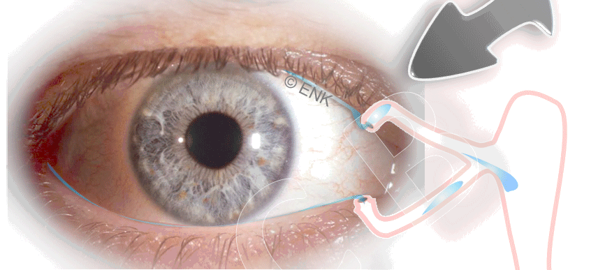
Eye and lacrimal drainage system. The tear drainage system begins on each lid at the tear point (lacrimal punctum), which is at the nasal side immersed into the tear lake (tear meniscus) to be able to suck up tear fluid. The lacrimal canaliculus runs from here within the edge of the eyelid towards the nose and both canaliculi unite in front of the lacrimal sac. The sack extends as the naso-lacrimal duct into the nose where the tears enter the nasal passage. If there is a very strong lack of aqueous tears, a punctum plug inside the tear point is useful to inhibit the removal of tear fluid from the eye. The pugging thus increases the height of the tear meniscus (´TMH´, please see in the animated image), which can be clinically measured as TMH-Test (=> here is more information with histological images)
If the formation of new tears is reduced, an obvious therapy option is to reduce the constant drainage of the already present tears from the ocular surface into the nose.
This will increase the amount of tears that remains on the eye and thus increase the height of tear meniscus (TMH).
The reduction of the tear outflow is achieved e.g. by so-called 'punctum plugs' (see animated figure). These are plastic plugs with which the lacrimal points can be temporarily closed - just like a plug in the sink.
Depending on the severity of the lack of tears, punctum plugs may be used in the lower eyelid only or also additionally in the upper eyelid:
with a plug in the lower eyelid (see animated figure), most of the tear flow is prevented - but some tears are still drained via the upper punctum
if a plug is also inserted into the upper eyelid, tear drainage is completely prevented.
Lacrimal tear punctum plugs / punctum plugs application
can be used very easily, quickly and painlessly at the slit lamp - this can be done with a small pair of tweezers or by a supplied applicator device.
can be removed just as easily, if required.
... occasionally they can even be unintentionally removed by intensive use of a washcloth or towel. But this is not the rule and depends on the type of plug and the size adjustment.
Punctum plugs used in the eyelids are typically not visible - that is, cosmetically harmless. They can only be seen in the considerable enlargement of the slit lamp.
Different models of punctum plugs
There are very different models of punctum plugs:
Material:
typically they consist of a more or less transparent plastic material that does not dissolve
but there are also materials that are supposed to dissolve after a predetermined, long period of time
Shape:
punctum plugs are usually shaped like a cone on one end so that they can be easily inserted into the lacrimal punctum
they typically have a flat part on top that covers the tear point
there are also models that are rod-shaped and inserted completely into the tear duct
a Punctum Plug typically has the task of completely preventing tears from flowing out. However, there are also models with a channel that only reduce tear drainage.
Surgical closure of the lacrimal punctum
If the treatment with plastic punctum plugs does not produce the desired result, or if the punctum plugs are frequently lost, the lacrimal puncta can also be closed surgically.
This can be done in the event of a very severe lack of tears and is in principle more permanent than punctum plugs.
This and other possible therapy options, such as anti-inflammatory medication, if necessary your own serum eye drops or surgical interventions, will be suggested by an experienced ophthalmologist or eye clinic.
